")
organisms. World-wide electronic publication, Kortenhoef, the Netherlands) for amoeboid organisms
or Encyclopedia of Life for all others.


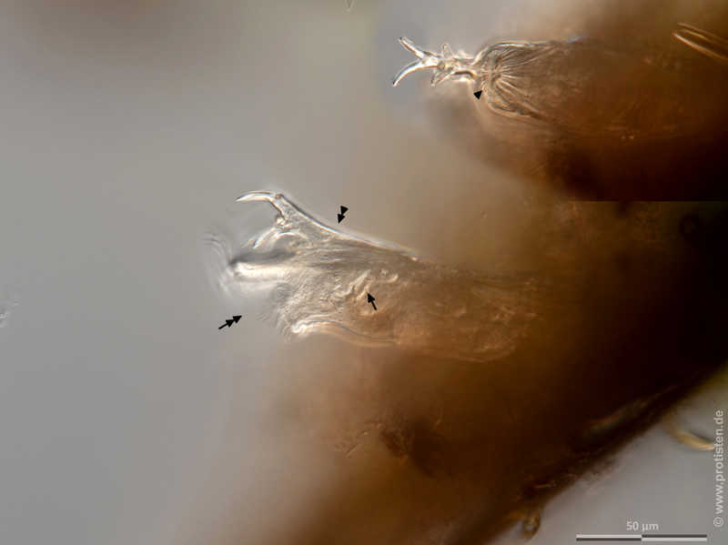
Ptygura kostei Fully expanded rotifer in lateral view in median optical section showing base rod and 3 of 4 hooks of the nuchal fork (double arrowhead), trophi (arrow) and fully opened corona. The inset above right shows the rotifer in ventral view with fully retracted somatic body, which allows an unimpeded view of the four hooks of the nuchal fork. Scale bar indicates 50 µm.
Sample from a tropical freshwater aquarium. Sampling date 3/2023. The image was built up using several photomicrographic frames with manual stacking technique. Images were taken using Zeiss Axioplan with Olympus OM-D M5 MKII.
Image under Creative Commons License V 3.0 (CC BY-NC-SA).
Place name: Tropical freshwater aquarium
Latitude: 54.3018013 Longitude: 10.07120132
Voll ausgestrecktes Rädertier in Seitenansicht im optischen Schnitt 3 von 4 Haken der Nackengabel (Doppelpfeilspitze), Kauer (Pfeil) und vollständig geöffneter Korona. Der Einschub oben rechts zeigt das Rädertierchen in ventraler Ansicht mit vollständig zurückgezogenem somatischem Körper, was einen unbehinderten Blick auf die vier Haken der Nackengabel ermöglicht. Multiebenen-Abbildung, manuell gestapelt. Der Messbalken markiert eine Länge von 50 µm.
Probe aus einem Süßwasseraquarium. Datum der Aufsammlung: 3/2023. Mikrotechnik: Zeiss Axioplan, Kamera: Olympus OM-D M5 MKII.
Creative Commons License V 3.0 (CC BY-NC-SA).
For permission to use of (high-resolution) images please contact postmaster@protisten.de.