")
organisms. World-wide electronic publication, Kortenhoef, the Netherlands) for amoeboid organisms
or Encyclopedia of Life for all others.


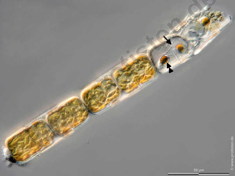
Melosira moniliformis The sexual reproduction of centric diatoms (Centrales) is characterized by oogamy. In Melosira, the spermatozoids are formed in narrower, the oogonia in broader filaments. After meiosis, four mobile spermatozoa with one flagellum develop in the male gamete mother cells (antheridia), in each of the oogonia three meiotic nuclei collaps and only one egg cell capable of fertilization remains.
The images show the flagella (arrow) and a golden brown chloroplast (double arrowhead, color due to the accessory dye fucoxanthin) at the posterior end of the spermatozoids (arrowhead).
After Krammer, K. and van den Hoek, C.
Scale bar indicates 50 µm. The specimen was gathered in the Kieler Förde (Baltic Sea). Sampling date 1/2022. Images were taken using Zeiss Axioplan with Olympus OM-D M5 MKII.
Image under Creative Commons License V 3.0 (CC BY-NC-SA).
Place name: Baltic Sea, Kieler Förde, Kiel Fjord (Germany)
Latitude: 54.3894126 Longitude: 10.1749055
Die sexuelle Fortpflanzung zentrischer Diatomeen (Centrales) zeichnet sich durch Oogamie aus. Bei Melosira werden die Spermien in schmaleren, die Oogonien in breiteren Zellketten gebildet. In den männlichen Gametenmutterzellen (Antheridien) entstehen nach der Meiose vier eingeißelige, bewegliche Spermatozioden, in den Oogonien sterben jeweils drei Meiose-Kerne ab und nur eine befruchtungsfähige Eizelle bleibt übrig.
Die Bilder zeigen die Geißel (Pfeil) sowie einen goldbraunen Chloroplasten (Doppelpfeilkopf, Farbe aufgrund des akzessorischen Farbstoffs Fucoxanthin) am Hinterende der Spermatozoiden (Pfeilkopf).
Nach Krammer, K. und van den Hoek, C.
Der Messbalken markiert eine Länge von 50 µm. Probe aus der Kieler Förde. Datum der Aufsammlung: 1/2022. Mikrotechnik: Zeiss Axioplan, Kamera: Olympus OM-D M5 MKII.
Creative Commons License V 3.0 (CC BY-NC-SA).
For permission to use of (high-resolution) images please contact postmaster@protisten.de.